![]() Figure 5 of
Pang, Mol Vis 2005;
11:152-162.
Figure 5 of
Pang, Mol Vis 2005;
11:152-162.
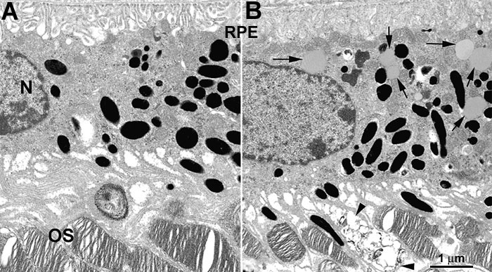
Figure 5. Electron microscopic changes in 3 month old retinas from normal C57BL/6J and rd12 mice
A: Normal RPE cells. B: rd12 RPE cells. No lipid-like droplets could be detected in normal RPE cells, while many typical lipid-like droplets (arrows) could be observed in the cytoplasm of rd12 RPE cells. Degenerating outer segments (OS; arrowheads) could be observed at this age. A nucleus (N) and the retinal pigment epithelium (RPE) are also labeled. The scale bar represents 1 μm.