![]() Figure 4 of
Joo, Mol Vis 2005;
11:133-142.
Figure 4 of
Joo, Mol Vis 2005;
11:133-142.
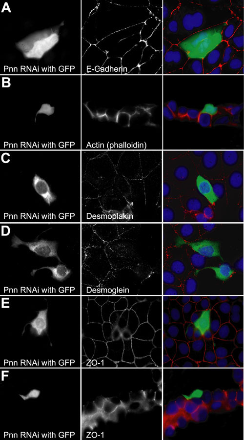
Figure 4. Levels of adherens, desmosomal, and tight junctional proteins are reduced after knock down of endogenous Pnn
HCE-T cells cotransfected with Pnn shRNAi and GFP vectors and immunostained with antibodies against E-cadherin, desmoplakin, desmoglein, and ZO-1 demonstrated significant reductions in cell-cell adhesion and junctional component staining. Staining for E-cadherin demonstrated large gaps along surfaces of Pnn shRNAi cells, which corresponded with cell processes that appeared to be crawling over the surface of neighboring cells (A). Cryostat sectioning of epithelial cells cultured on membrane inserts revealed that the GFP/Pnn-RNAi cells leave the plane of the epithelia and seem to reside on top of the confluent sheet (B). Desmoplakin (C), desmoglein (D), and ZO-1 (E,F) all exhibit reduced staining in the GFP/PnnRNAi cells with the presence of extensive cellular processes that interdigitate and/or crawl on the surface of the epithelial sheets. Cryostat sectioning of transfected epithelial cells clearly demonstrate Pnn-RNAi cells above the plane of the tight junction as shown by ZO-1 immunostaining (F).