![]() Figure 2 of
Joo, Mol Vis 2005;
11:133-142.
Figure 2 of
Joo, Mol Vis 2005;
11:133-142.
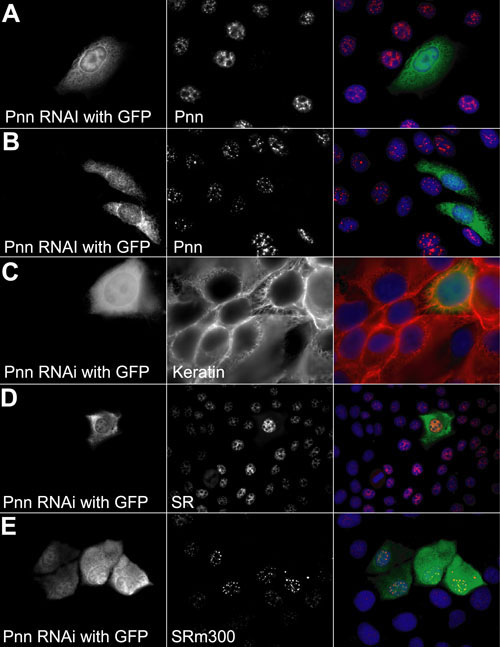
Figure 2. Knock down of endogenous Pnn protein levels by Pnn-RNAi reduces cell-cell adhesion and alters distribution of Pnn associated components
HCE-T cells were transfected with Pnn shRNAi and GFP vectors. After 24 h (A) and 48 h (B), cells were fixed in methanol and immunostained with Pnn-antibody. The GFP positive cells exhibited a clear knockdown in Pnn expression. In addition, the cell shape and relationship of the GFP/Pnn-RNAi cells with neighboring cells were altered. The GFP positive cell exhibited separation from surrounding cells and a more elongate morphology. Keratin filaments appeared to lose their membrane anchorage and condense in the perinuclear region in the GFP/Pnn-RNAi cells (C). Knocking down Pnn was associated with the accumulation of SR proteins, notably SRm300 in larger nuclear speckles (D,E).