![]() Figure 1 of
Joo, Mol Vis 2005;
11:133-142.
Figure 1 of
Joo, Mol Vis 2005;
11:133-142.
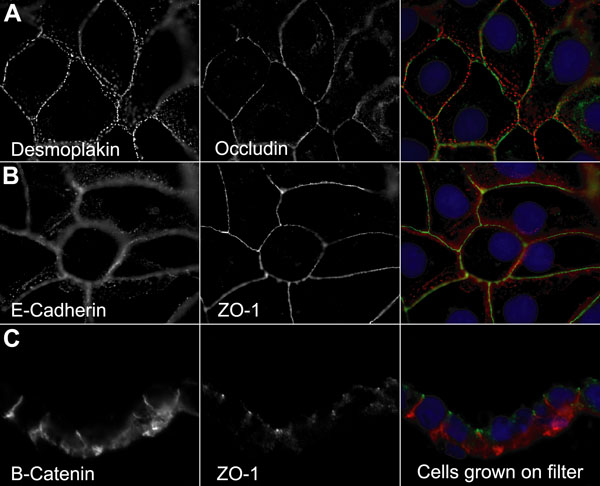
Figure 1. HCE-T cells form extensive intercellular junctions and polarized epithelia
HCE-T cells show epithelial polarization with E-cadherin and desmosome (desmoplakin staining) distributed throughout the lateral surface, while tight junction components were restricted to the most apical aspect of the lateral cell-cell contacts. Cells were grown to confluency on coverslips and immunostained with antibodies against desmoplakin (red), E-cadherin (red), occludin (green), and ZO-1 (green; A,B). Cell nuclei were visualized with DAPI (blue). Epithelial monolayers grown on filter inserts were also sectioned and immunostained with β-catenin (red) and ZO-1 (green) to demonstrate HCE-T epithelial polarization with lateral surface staining of β-catenin and the apically placed ZO-1 (C).