![]() Figure 8 of
Rex, Mol Vis 2005;
11:1236-1245.
Figure 8 of
Rex, Mol Vis 2005;
11:1236-1245.
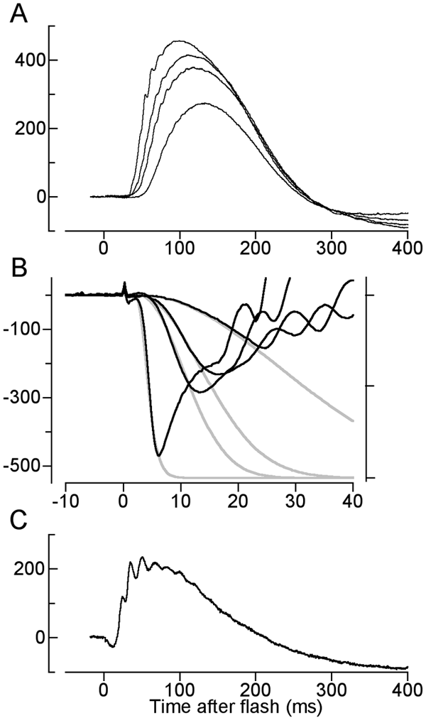
Figure 8. Electroretinographic recordings from a mouse expressing EGFP under control of the b-actin promoter
A: Scotopic b-waves are responses to ganzfeld flashes of luminance 3x10⁻⁴, 8.6x10⁻³, and 3.2x10⁻³ scotopic cd s m-2. B: Rod a-waves are responses to flashes of 0.7, 3.7, 7.5, and 206 scotopic cd s m-2. The traces are shown on a ten fold faster time base than those in A; the saturating amplitude, obtained in response to the most intense flash is 471 mV. The normalized traces (right ordinate) have been fitted with a model of the rod phototransduction cascade [33]. Assuming a dilated pupil area of 3.2 mm2 and conversion factor for scot Td s to photoisomerizations of 171 [34], the amplification coefficient obtained from the fitting is A=4.4 s-2. C: Photopic b-wave are the same intensity flash as was used to produce the saturating a-wave in B was presented in the presence of a steady background that suppressed rod activity (as evidenced by the absence of all but a small a-wave, which is attributable to cones). The cone-driven b-wave amplitude (with oscillations filtered out) is 200 mV.