![]() Figure 6 of
Rex, Mol Vis 2005;
11:1236-1245.
Figure 6 of
Rex, Mol Vis 2005;
11:1236-1245.
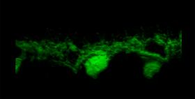
Figure 6. Morphology of a horizontal cell from an AAV2/5.CMV.EGFP-infected retina
Three-dimensional representation of a z-stack of confocal images from an AAV2/5.CMV.EGFP infected mouse retina showing the distribution of enhanced green fluorescent protein in a representative horizontal cell.
Note that the slide bar at the bottom of the quicktime movie can be used to manually control the flow of the movie. A representative frame from the movie is included below.
| This animation requires Quicktime 6 or later. Quicktime is available as a free download. |