![]() Figure 5 of
Rex, Mol Vis 2005;
11:1236-1245.
Figure 5 of
Rex, Mol Vis 2005;
11:1236-1245.
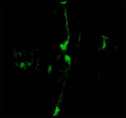
Figure 5. Morphology of a Müller cell from an AAV2/5.CMV.EGFP-infected retina
Three-dimensional confocal images showing distribution of enhanced green fluorescent protein in a Müller cell. One Müller cell is seen to span the entire width of the retina in this image and portions of others are apparent as well. For the cell which spans the entire retina, the endfoot (vitreal aspect of the cell) is oriented toward the top of the image and the portion of the cell adjacent to photoreceptors is at the bottom of the image. Müller cell nuclei appear as rounded structures approximately mid-way across the retina. Processes project from the main trunks of the Müller cells.
Note that the slide bar at the bottom of the quicktime movie can be used to manually control the flow of the movie. A representative frame from the movie is included below.
| This animation requires Quicktime 6 or later. Quicktime is available as a free download. |