![]() Figure 1 of
Rex, Mol Vis 2005;
11:1236-1245.
Figure 1 of
Rex, Mol Vis 2005;
11:1236-1245.
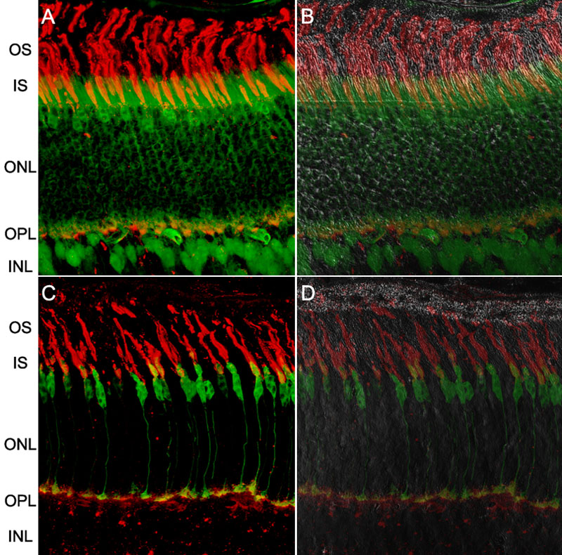
Figure 1. Distribution of enhanced green fluorescent protein in photoreceptors in transgenic mice
The retinal structure of mice expressing enhanced green fluorescent protein (EGFP) under two different promoters-one constitutive and the other cell-specific, is typical of that found normally in a C57Bl/6 wild type (WT) retina. A,B: Images of a frozen section of a retina of a mouse expressing EGFP under control of the β-actin promoter. C,D: Images of a section of the retina of a mouse expressing EGFP under control of the human cone L/M opsin promoter. A,C: Confocal fluorescence scans with the green channel reporting EGFP fluorescence, while the red channel shows PNA staining, primarily of the cone matrix sheaths. B,D: Differential interference contrast (DIC) images overlaid on the corresponding fluorescence sections at left, with the DIC images shown at 65% transparency. The images are each about 100 μm x 100 μm. The outer segment layer (OS), inner segment layer (IS), outer nuclear layer (ONL), and outer plexiform layer (OPL) are identified.