![]() Figure 6 of
Bejjani, Mol Vis 2005;
11:124-132.
Figure 6 of
Bejjani, Mol Vis 2005;
11:124-132.
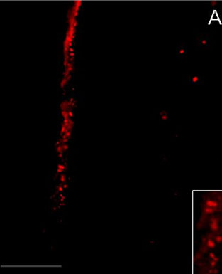
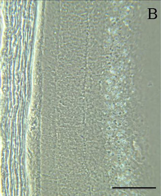
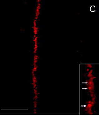
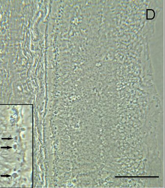
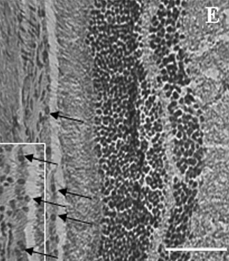
Figure 6. In vivo gene expression of red nuclear fluorescent protein within RPE cells after intravitreal injection.
Phase contrast and fluorescent microscopy of rat retina sections after intravitreous injection of nanoparticles loaded with RNFP plasmid: seven days (A,B) and fourteen days (C,D) after the initial intravitreal injection of nanoparticles. Inserts demonstrate the localization of the red fluorescence (expression of RNFP) within the RPE nuclei. E: Hematoxylin-eosin stained rat retina section. Arrows show nuclei. Bars represent 20 μm.