![]() Figure 2 of
Bejjani, Mol Vis 2005;
11:124-132.
Figure 2 of
Bejjani, Mol Vis 2005;
11:124-132.
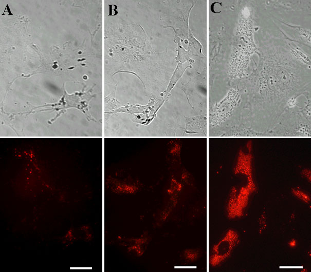
Figure 2. Kinetics of nanoparticle internalization by bovine RPE cells
Phase and fluorescence microscopy of RPE cells incubated with 1 mg/ml NP-Rh. Removing the added nanoparticles (1.0 mg/ml) after 1 h of incubation and extending the culture for 72 h shows that each RPE cell in culture harbors a few NPs in its cytoplasm. The number of internalized NPs per cell increases rapidly when the contact time between cultured RPE cells and NPs is increased up to 6 h. Extended NP incubation times do not further increase internalization. A: Incubated 1 h. B: Incubated 3 h. C: Incubated 6 h. Bars represent 20 μm.