![]() Figure 1 of
Bejjani, Mol Vis 2005;
11:124-132.
Figure 1 of
Bejjani, Mol Vis 2005;
11:124-132.
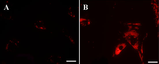
Figure 1. Fluorescence microphotographs of bovine RPE cells incubated with fluorescent nanaoparticles
Fluorescence microphototgraphs of bovine RPE cells incubated with fluorescent nanaoparticles for 6 h, showing that intracellular content of nanoparticles depend on their concentration in the medium. RPE cells incubated with 0.1 mg/ml (A) and 4 mg/ml (B) of NP-Rh for 6 h. Ingested nanoparticles appear as red fluorescent dots. Bars represent 20 μm.