![]() Figure 6 of
Shimauchi-Matsukawa, Mol Vis 2005;
11:1220-1228.
Figure 6 of
Shimauchi-Matsukawa, Mol Vis 2005;
11:1220-1228.
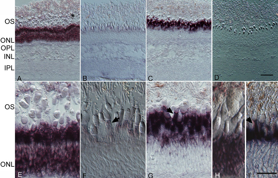
Figure 6. Retinal expression pattern of carp GRK genes
A-C,E-G: Antisense cRNA probes were hybridized to detect expression of GRK1A-1a (A,E), GRK1Ba (B,F), GRK7-1a (C,G), rhodopsin (H) and red sensitive cone pigment (I). D: Sense cRNA probe was used for a negative control of GRK7-1a. A-D: Low magnification. E-I: high magnification. Scale bars represent 50 μm in A-D and 20 μm in E-I. The expression of GRK1A-1a and rhodopsin was found throughout the outer nuclear layer, and the expression of GRK1B, GRK7-1a, and red sensitive pigment was observed in the myoid region just below the cone ellipsoid (arrows).