![]() Figure 5 of
Nguyen, Mol Vis 2005;
11:1183-1199.
Figure 5 of
Nguyen, Mol Vis 2005;
11:1183-1199.
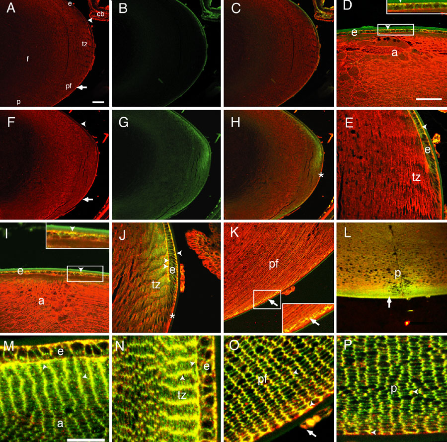
Figure 5. Co-localization of Scrib with E- and N-cadherin in the lens
To determine if Scrib co-localized with E- or N-cadherin in the epithelial and fiber cells of the lens, we conducted double immunofluorescence experiments on eye sections from P10 mice. Staining for Scrib and E- and N-cadherin overlapped on the basal surface of he epithelium. Staining for Scrib and N-cadherin overlapped extensively on the short sides of the fibers at the epithelial-fiber interface and on and on the basolateral surfaces of the fiber cells in the posterior of the lens. The figure shows immunofluorscent staining in the lens for Scrib only (A,F), E-cadherin only (B), N-cadherin only (G), Scrib and E-cadherin (C,D,E), and Scrib, and N-cadherin (H-P). A-L are longitudinal sections and M-P are transverse sections showing the regions corresponding to those in I-L. A-C,F-H are at low magnification; D-E,I-L are at intermediate magnifications; and M-P are at high magnification. Staining for Scrib is red, staining for E-cadherin, N-cadherin and ZO-1 is green, and overlap in staining for Scrib and E-cadherin, N-cadherin or ZO-1 is yellow. The arrowheads (A,F) mark the basal surfaces of epithelial cells. The arrows in A,F show the strong Scrib staining in basal tips of fiber cells. The asterisks (H,J) show the absence of staining for N-cadherin along the capsule in the posterior transition zone. The arrowhead in D shows overlap in staining for Scrib and E-cadherin in the basal membranes of the epithelium. The arrowhead in E shows overlap in staining for Scrib and E-cadherin on the basal cell surface of the epithelial cells in the transition zone. The arrowheads in I,J show overlap of staining for Scrib and N-cadherin along the basal surfaces of the epithelial cells (I) and the basal surfaces of the cells of the transition zone (tz; J) while the double arrowheads in J show overlap in staining at the apical interface between the epithelium and fiber cells. The arrow in K shows the overlap in staining along basal and lateral surfaces in the fiber cells and the arrow in L shows the staining overlap of Scrib and N-cadherin in the posterior of the lens. The insets in D,I,K show the detail of overlapping staining patterns in the regions corresponding to the areas in white boxes in the respective panels. The insets in D,I, arrowheads, show in detail the overlap in staining for Scrib and E- and N-cadherin on the basal surface of the epithelium, respectively. The inset in K, arrow, shows the detail of overlap in Scrib and N-cadherin staining on the lateral membranes just off the basal surface in the posterior fiber region. In M,N, the arrowheads show overlap (yellow) in staining of Scrib and N-cadherin on the short sides of the fiber cells and at the epithelial-fiber interface. In O,P, the arrowheads show overlap in staining of Scrib and N-cadherin on the short sides of the fiber cells and the basal attachments of the fiber cells to the capsule. Note that with the exception of the extreme basal region, Scrib is restricted to the short sides of the fibers while N-cadherin, although more concentrated on the short sides, is found on both. The arrow in D shows Scrib staining in the blood vessels outside the lens capsule. The anterior (apical) region of fiber cells (a), ciliary body (cb), lens epithelium (e), lens fiber cells (f), midposterior fibers (pf), and posterior (basal) region of the lens fibers (p) are identified. In all panels, the anterior the eye is oriented at the top. The scale bars represent 100 μm in A-C,F-H; 50 μm in D,E,I-L; 25 μm in the insets in D,I,K; and 25 μm in M-P.