![]() Figure 4 of
Nguyen, Mol Vis 2005;
11:1183-1199.
Figure 4 of
Nguyen, Mol Vis 2005;
11:1183-1199.
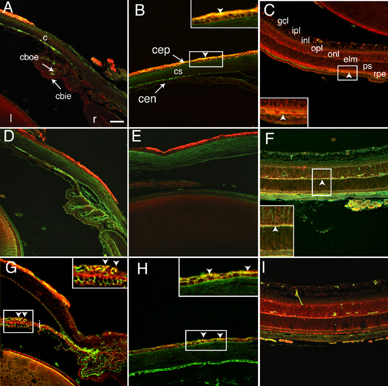
Figure 4. Co-localization of Dlg-1 and adhesion proteins in the eye
To determine if Dlg-1 co-localized with E- or N-cadherin throughout the eye, we conducted double immunofluorescence experiments on eye sections from P10 mice. Staining for Dlg-1 overlapped with E-cadherin in the corneal epithelium and RPE, with N-cadherin in the neural retina and with ZO-1 in the corneal epithelium and iris. The figure shows immunofluorescent staining in the cornea (c; A,H), iris (i) and ciliary body (cb; A,D,E,G) and retina (r; C,F,I) for Dlg-1 and E-cadherin (A-C), Dlg-1 and N-cadherin (D-F), and Dlg-1 and ZO-1 (C-I). Staining for Dlg-1 is red, staining for E-cadherin, N-cadherin and ZO-1 is green and overlap in staining for Dlg-1 and E-, cadherin, N-cadherin, or ZO-1 is yellow. The insets in B,C,F,H show the detail of overlapping staining patterns in the regions corresponding to the areas in white boxes in their respective panels. The insets in B,C, arrowheads, show the overlap in staining for Dlg-1 and E-cadherin in the corneal epithelium (cep), and retinal pigment epithelium (rpe) of the retina, respectively. The inset in F, arrowhead, shows the overlap in staining for Dlg-1 and N-cadherin in the external limiting membrane (elm) of the retina. The inset in G, arrowheads, shows the overlap in staining of Dlg-1 and ZO-1 in the iris. The inset in H, arrowheads, shows overlap in staining for Dlg-1 and ZO-1 in the corneal epithelium. The ciliary body outer epithelium (cboe), ciliary body inner epithelium (cbie), lens (l), corneal epithelium (cep), corneal stroma (cs), corneal endothelium (cen), ganglion cell layer (gcl), inner plexiform layer (ipl), inner nuclear layer (inl), outer plexiform layer (opl), outer nuclear layer (onl), and outer and inner segments of photoreceptors (ps) are identified. In all panels, the anterior of the eye is oriented at the top. The scale bar represents 50 μm in all panels and 25 μm for the insets in B,C,F,H.