![]() Figure 1 of
Nguyen, Mol Vis 2005;
11:1183-1199.
Figure 1 of
Nguyen, Mol Vis 2005;
11:1183-1199.
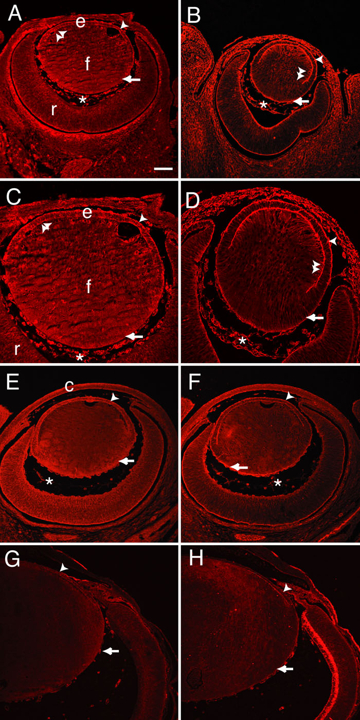
Figure 1. Expression of Dlg-1 and Scrib in eye development
To determine where Dlg-1 and Scrib were found in the eye during embryonic development, we conducted immunofluorescence experiments on eye sections from embryos of various ages and from neonates. Shown are immunostaining for Dlg-1 (A,C,E,G) and Scrib (B,D,F,H); at E13.5 (A-D), E15.5 (E,F), neonate (G,H). C,D are higher magnifications of the sections in A,B, respectively. Dlg-1 and Scrib staining was observed in the epithelium (e) and fiber (f) cells of the lens, cornea (c), and retina (r) throughout early development of the eye. The arrowheads in A,C,E,G show the basal surface of the epithelium where strong Dlg-1 staining was frequently observed. The double arrowheads in A,C show strong staining for Dlg-1 at the epithelial-fiber interface. The arrows in A,C,E,G show staining for Dlg-1 in basal regions of the lens fiber cell compartment. The arrowheads in B,D,F,H show strong staining for Scrib in the basal membranes of the epithelium. The double arrowheads in B,D show strong staining for Scrib in the epithelial-fiber junction and in the apical regions of the fiber cells. The arrows in B,D,F,H show strong Scrib staining in the basal surface of the fiber cell compartment. The asterisks in A-F show staining in the hyaloid vessels. lens epithelium (e), cornea (c). In all panels, the anterior of the eye at the top. The scale bar represents 100 μm for A,B,E-H and 50 μm for C,D.