![]() Figure 7 of
Hemati, Mol Vis 2005;
11:1151-1165.
Figure 7 of
Hemati, Mol Vis 2005;
11:1151-1165.
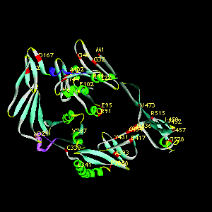
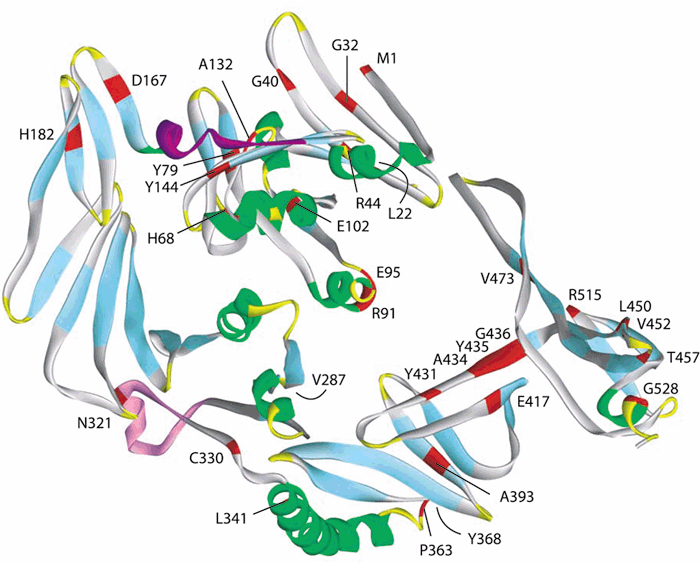
Figure 7. Ab initio model of RPE65 tertiary structure
A: A three-dimensional structure for the RPE65 protein was predicted using the method of Bystroff and Shao [39]. The ribbon representing the peptide backbone is color coded according to structural components: α-helices are green; β-pleated sheets are blue; and random coil is gray. The linear sequence corresponding to the epitope recognized by mAb 8B11 is shown in purple and for mAb 1F9 is shown in pink. Sites of amino acid substitutions resulting from patient missense mutations associated with inherited retinal degeneration in patients are shown in red. B: The structure is animated and rotates about the vertical axis.
Note that the slide bar at the bottom of the movie can be used to manually control the flow of the movie. If you are unable to view the movie, a representative frame is included below.
A:
B:
| This animation requires Quicktime 6 or later. Quicktime is available as a free download. |