![]() Figure 11 of
Hemati, Mol Vis 2005;
11:1151-1165.
Figure 11 of
Hemati, Mol Vis 2005;
11:1151-1165.
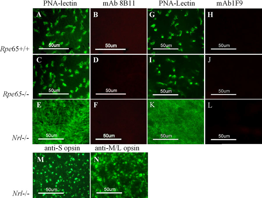
Figure 11. Immunohistochemical analysis of retina flatmounts
Retinas from wild type (A,B,G,H), Rpe65 knockout (C,D,I,J), and Nrl knockout mouse (E,F,K-N) were fixed on coverslips and incubated with PNA-lectin (FITC-conjugated), mAb 8B11, mAb 1F9, or antibodies against S-opsin and M/L-opsin. Alexa Fluor 555-conjugated anti-mouse IgG was used as secondary antibody. PNA labeling (A,C,E,G,I,K), mAb 8B11 labeling (B,D,F), mAb 1F9 labeling (H,J,L), S-opsin labeling (M), M/L-opsin labeling, (N). Fluorescence imaging: PNA-lectin, opsin antibodies 1/60 s; mAb 8B11 1/12 s; mAb 1F9, 1/20 s. The scale bars represent 50 μm.