![]() Figure 10 of
Hemati, Mol Vis 2005;
11:1151-1165.
Figure 10 of
Hemati, Mol Vis 2005;
11:1151-1165.
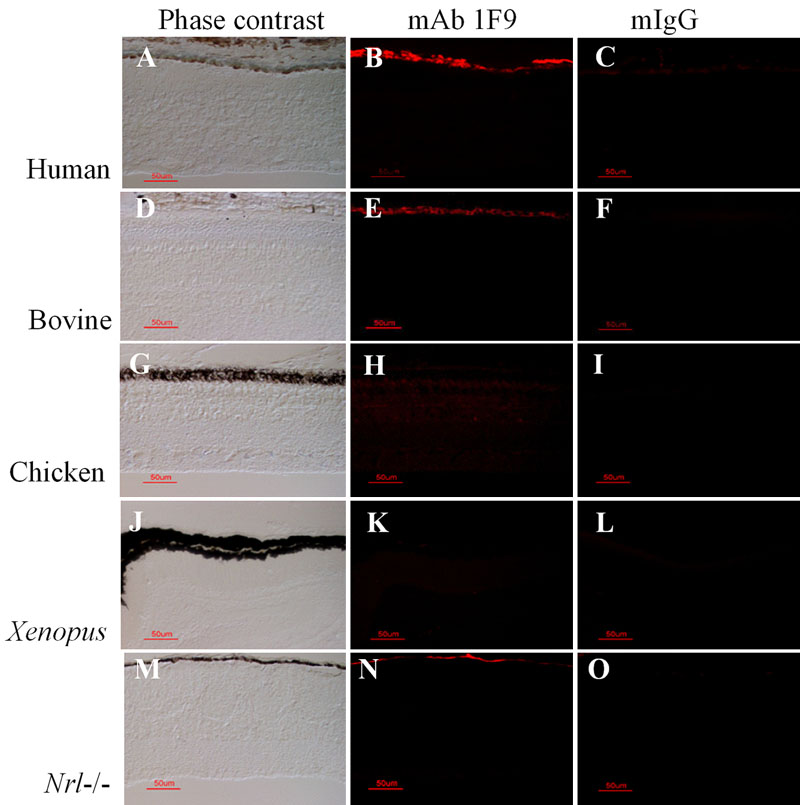
Figure 10. Immunohistochemical analysis of mAb 1F9 reactivity in rod- and cone-dominant retinas
Retina/choroid/RPE cryosections of paraformaldehyde-fixed eyes from bovine, human (rod-dominant), and from Xenopus laevis, chicken, Nrl knockout mouse (cone-dominant) were incubated with mAb 1F9 or mouse non-immune IgG using M.O.M. Peroxidase reagents, and visualized using TSA-Alexa fluor 568 reagents using fluorescence imaging (1/50 s human, all others 1/30 s). Phase contrast (A,D,G,J,M), mAb 1F9 reactivity (B,E,H,K,N), non-immune mouse IgG reactivity (C,F,I,L,O). Bovine sections are from an amelanotic region of the RPE. The scale bars represent 50 μm.