![]() Figure 1 of
Hemati, Mol Vis 2005;
11:1151-1165.
Figure 1 of
Hemati, Mol Vis 2005;
11:1151-1165.
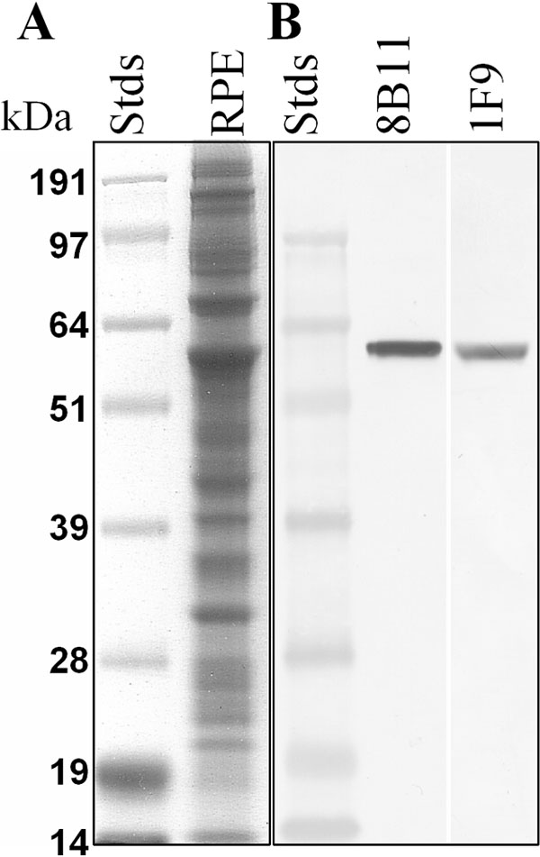
Figure 1. Immunoreactivity of mAb 8B11 and mAb 1F9 with bovine RPE membranes
A: Coomassie blue staining of proteins separated by SDS-PAGE (20 μg protein). B: Western analysis (1 μg protein) of proteins separated by SDS-PAGE, transferred to nitrocellulose, incubated with mAb 811 (0.2 μg/ml), or mAb 1F9 (3 μg/ml), and immunoreactivity visualized using alkaline phosphatase coupled anti-mouse IgG. RPE65 migrates at about 61 kDa.