![]() Figure 7 of
Warburton, Mol Vis 2005;
11:1122-1134.
Figure 7 of
Warburton, Mol Vis 2005;
11:1122-1134.
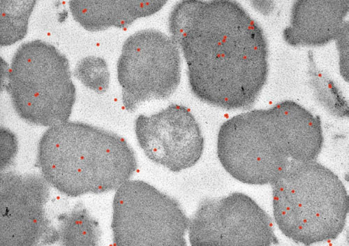
Figure 7. Immunocyctochemistry of RLF
RLF granules were fixed and embedded in resin. Thin sections were collected on gold grids, incubated with an anti-rhodopsin antibody, washed, incubated with gold-conjugated protein A, washed, and examined using transmission electron microscopy (TEM). TEM showed gold-conjugated protein A bound to anti-rhodopsin antibodies on the RLF granules as dark black dots, which have been pseudocolored red. Rhodopsin is an integral part of the RLF granules.