![]() Figure 6 of
Warburton, Mol Vis 2005;
11:1122-1134.
Figure 6 of
Warburton, Mol Vis 2005;
11:1122-1134.
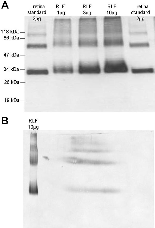
Figure 6. Rhodopsin immunoblots of RLF
A: Immunoblot following SDS-PAGE of 1, 3, and 10 μg of total RLF protein. Shown for comparison is 2 μg of protein from photoreceptor cell membranes enriched from human retina. Rhodopsin runs on SDS-PAGE as a mixture of the monomer (about 30 kDa), dimer (about 60 kDa), and trimer (about 90 kDa). B: Immunoblot following 2D electrophoresis of 10 μg of total RLF protein. For comparison, 10 μg of RLF protein was loaded in the standards lane of the second dimension gel. The diffusion (band broadening, due to modifications) of the rhodopsin in RLF as compared with that in photoreceptor cells indicates that the rhodopsin in RLF is remarkably heterogeneous.