![]() Figure 5 of
Warburton, Mol Vis 2005;
11:1122-1134.
Figure 5 of
Warburton, Mol Vis 2005;
11:1122-1134.
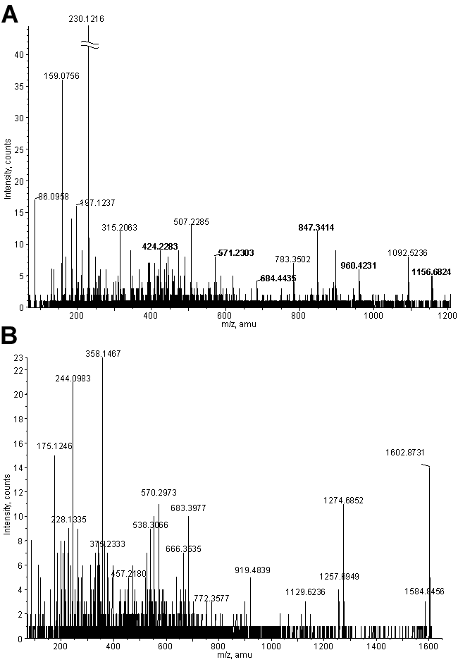
Figure 5. Mass spectrometry of peptides from RLF proteins
A: CID spectrum from LCMSMS of the peptide identified as SAAIYNPVIYIMO2MONK (where MO represents Met sulfoxide and MO2 represents Met sulfone) from rhodopsin. Indicated in bold are six y ions that make identification of these modified sites unequivocal. Another spectrum showed the same peptide with Met sulfoxide at the first Met position, and a third spectrum showed Met sulfone at the first Met without Met sulfoxide at the second Met position (data not shown). B: Representative CID spectrum from oMALDI MSMS. This peptide was identified as LVDQNIFSFYLSR from cathepsin D. The spectrum in A allows the conclusive identification of the modified methionines at positions 308 and 309 of rhodopsin. These spectra show that the fragmentation data obtained by both LCMSMS and oMALDI MSMS are of sufficiently high quality to allow unambiguous identification of the corresponding peptides.