![]() Figure 3 of
Warburton, Mol Vis 2005;
11:1122-1134.
Figure 3 of
Warburton, Mol Vis 2005;
11:1122-1134.
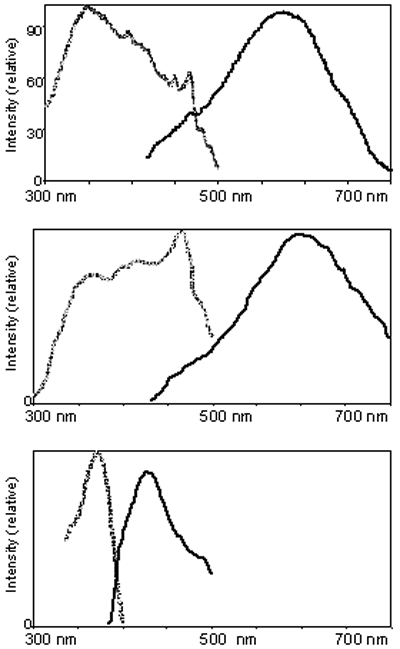
Figure 3. Fluorescence of RLF granules
Fluorescence spectroscopy of RLF from our preparations (top panel) compared with RLF from Boulton et al. [12] (middle panel) and Schutt et al. [11] (lower panel). The spectra from their published papers were simply traced. Excitation (gray) monitored with emission at 570 nm, and emission (black) monitored with excitation at 364 nm. Our evidence that supports the congruence of our preparations with those studied by Boulton et al. [12].
