![]() Figure 4 of
Zinchuk, Mol Vis 2005;
11:114-123.
Figure 4 of
Zinchuk, Mol Vis 2005;
11:114-123.
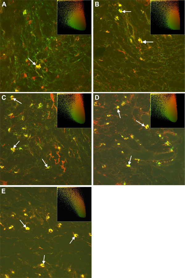
Figure 4. Double staining for PAF-R and MBP antigens
Confocal immunofluorescence microscopy of conjunctiva sections stained with anti-PAF-R (red fluorescence) and anti-MBP (green fluorescence) antibodies. The embedded scatter gram in the upper right corner of each image estimates the amount of each detected antigen based on colocalization of PAF-R (red, y-axis) and MBP (green, x-axis). Colocalized pixels of yellow color are located along the diagonal of the scatter gram. All images reveal colocalization of PAF-R and MBP antigens at different time points (arrows, A-E). Magnification x400.