![]() Figure 3 of
Zinchuk, Mol Vis 2005;
11:114-123.
Figure 3 of
Zinchuk, Mol Vis 2005;
11:114-123.
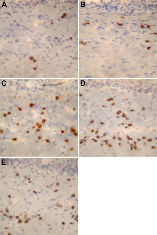
Figure 3. Immunohistochemical visualization of MBP antigen
MBP positive cells are seen occasionally in controls (A). The number of MBP positive cells more than doubles at 30 min after PAF challenge (B). It increases dramatically at 2 h (C) and keeps increasing until 6 h (D). At 24 h, a decrease in the number of immunopositive cells is noticed (E). Representative images of three examined eyes are shown. Magnification x400.