![]() Figure 2 of
Zinchuk, Mol Vis 2005;
11:114-123.
Figure 2 of
Zinchuk, Mol Vis 2005;
11:114-123.
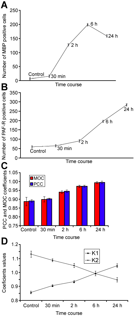
Figure 2. Dynamical changes of the number of PAF-R and MBP positive cells and colocalization parameters
Quantitative representation of the changes of the number of PAF-R and MBP immunopositive cells (A and B), Manders' colocalization and Pearson's correlation coefficients (C), and overlap coefficients K1 and K2 (D) in dynamics of PAF induced conjunctivitis. A and B document an increase of the number of MBP and PAF-R positive cells with their respective peaks at 6 and 24 h after PAF administration. Values at each time point were compared to values at all other time points. All of these comparisons were found to be statistically significant (p<0.01, Mann-Whitney U test). C and D reveal changes of colocalization of PAF-R and MBP antigens. Manders' Overlap Coefficients (MOC) and Pearson's correlation coefficients (PCC) indicate a steady increase of the degree of colocalization during the whole period of observation (C), while overlap coefficients K1 and K2 indicate profound changes of the contribution of PAF-R and MBP antigens to their colocalization (D). Values at each time point were compared to values at all other time points. All of these comparisons were found to be statistically significant (p<0.05, Mann-Whitney U test). Data are an average of three samples examined for each time point.