![]() Figure 7 of
Liu, Mol Vis 2005;
11:1112-1121.
Figure 7 of
Liu, Mol Vis 2005;
11:1112-1121.
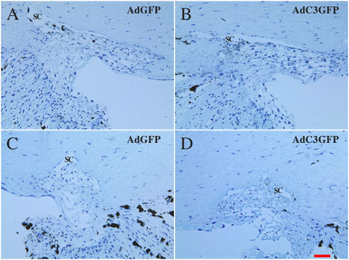
Figure 7. Morphology of the trabecular meshwork in organ cultured monkey anterior segments
Anterior segments were transduced with AdGFP (A,C) or AdC3GFP (B,D). Light microscopy of transduced anterior segments shows that changes in morphology did not correspond with the vector used. Compared to the control anterior segments (A,C), those transduced with the AdC3GFP vector exhibited either the same degree of cellularity and organization of the beams (B) or showed a decrease in cellularity (D). Schlemm's canal (SC) is identified. The scale bar represents 50 μm.