![]() Figure 2 of
Liu, Mol Vis 2005;
11:1112-1121.
Figure 2 of
Liu, Mol Vis 2005;
11:1112-1121.
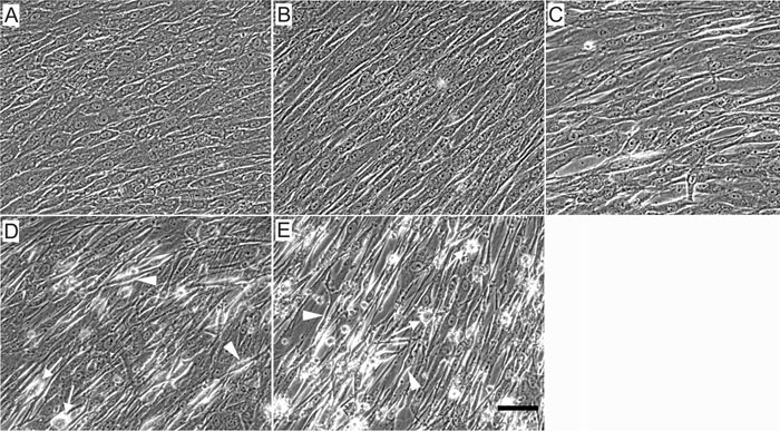
Figure 2. Human trabecular meshwork cell morphology after transduction with adenovirus vectors
Human trabecular meshwork cells were transduced with AdGFP or AdC3GFP for 4 days. A: Control, nontransduced cells. B,C: Control cells transduced with AdGFP at MOIs of 2.5 and 25 appeared slightly different in morphology and were arranged in a more random orientation. D,E: Cells transduced with AdC3GFP at MOIs of 2.5 and 25 appeared to be either partially retracted, rounded up completely (arrows), or very elongated (arrowheads) compared to control cells. No obvious cell detachment was observed in any cells. The scale bar represents 50 μm.