![]() Figure 5 of
Berube, Mol Vis 2005;
11:1101-1111.
Figure 5 of
Berube, Mol Vis 2005;
11:1101-1111.
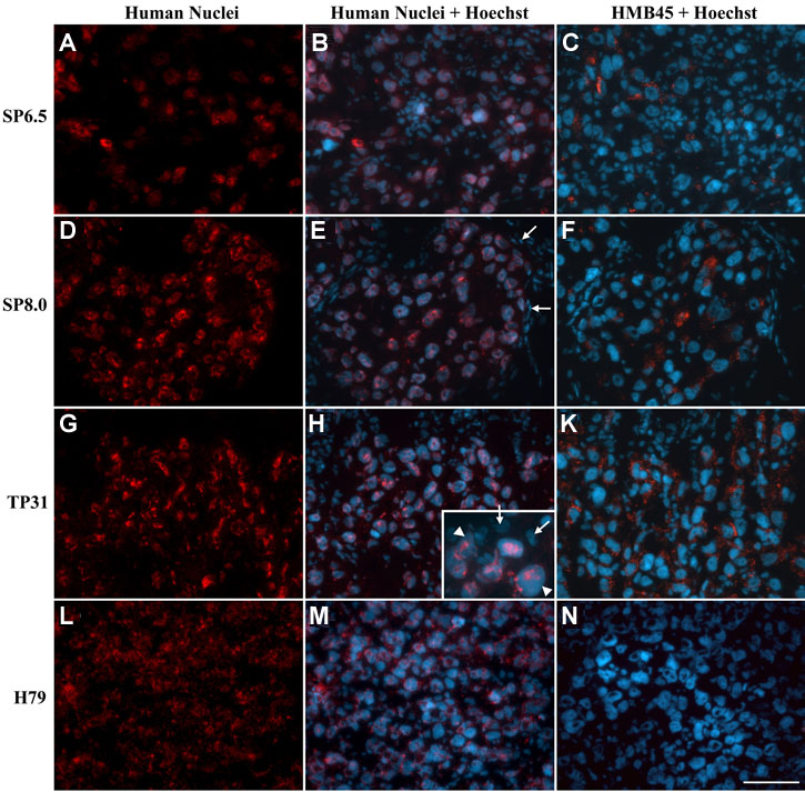
Figure 5. HMB45 expression and human nuclei staining in uveal melanoma-derived tumors
A,D,G,J: Human nuclear staining (red) of 5 μm thick paraformaldehyde-fixed cryosections. B,E,H,K: Human nuclear staining (red) merged with Hoechst staining (blue) of all cell nuclei (panel H insert is a magnification of H). C,F,I,L: HMB45 staining (red) on consecutive cryosections of the tumors. Cell nuclei were counterstained with Hoechst (blue). Scale bar represents 50 μm. E: Arrows identify cells originating from the chick embryos. H: Arrows and arrowheads in indicate the position of chicken and human cells, respectively.