![]() Figure 4 of
Berube, Mol Vis 2005;
11:1101-1111.
Figure 4 of
Berube, Mol Vis 2005;
11:1101-1111.
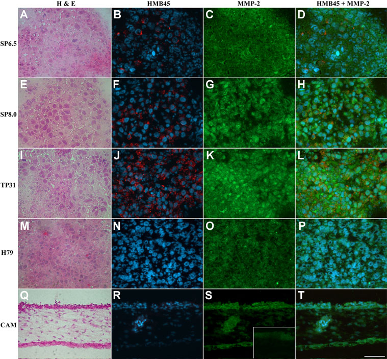
Figure 4. HMB45 and MMP-2 expression in human uveal melanoma-derived tumors
A,E,I,M,Q: Hematoxylin and eosin staining of 5 μm thick ethanol/formol-fixed cryosections. Panels B,F,J,N,R: HMB45 staining (red) on consecutive paraformaldehyde-fixed cryosections of the tumors. C,G,K,O,S: MMP-2 staining (green) of the same cryosections as those stained with the HMB45 Ab (the S insert is a negative control for S). D,H,L,P,T: Merge of HMB45 (red) and MMP-2 (green) stainings. Cell nuclei were counterstained with Hoechst (blue). The scale bar represents 50 μm.