![]() Figure 2 of
Berube, Mol Vis 2005;
11:1101-1111.
Figure 2 of
Berube, Mol Vis 2005;
11:1101-1111.
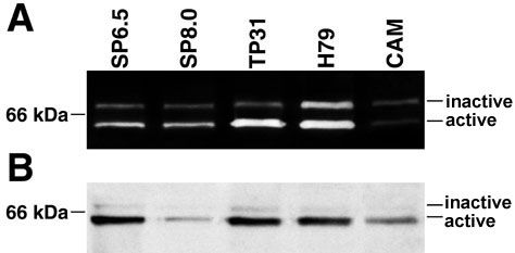
Figure 2. Zymographic and western blot analysis of MMP-2 from the in vivo tumors
A: Representative zymographic profiles of gelatinase activities in tumor tissues. The in vivo gelatinolytic activity of MMP-2 was determined by zymographic analysis of pro- and active MMP-2 in tissue extracts (50 μg each) from the tumors generated by the inoculation of the SP6.5, SP8.0, TP31, and H79 uveal melanoma cell lines on the CAM of chick embryos. The 72 kDa proform and the 62 kDa activated form of MMP-2 are indicated (inactive and active forms of MMP-2, respectively). B: Western blot analysis of MMP-2 in chick embryo's tumors produced by the uveal melanoma cell lines. For each sample, equal amounts of proteins (50 μg) were loaded on a 10% SDS-polyacrylamide gel prior to their transfer onto the membrane and further analysis with a monoclonal Ab directed against MMP-2. The position of the 66 kDa molecular mass marker is provided. C: Activation ratios of MMP-2 in the chick embryo tumor tissues derived from the uveal melanoma cell lines. The activation ratios were measured by computer-assisted image analyses of the gels. Horizontal bars indicate the mean values for each group. The error bars represent the standard error of the mean of the activation ratios of MMP-2. Triple asterisks indicate p<0.0001 compared to CAM.