![]() Figure 1 of
Berube, Mol Vis 2005;
11:1101-1111.
Figure 1 of
Berube, Mol Vis 2005;
11:1101-1111.
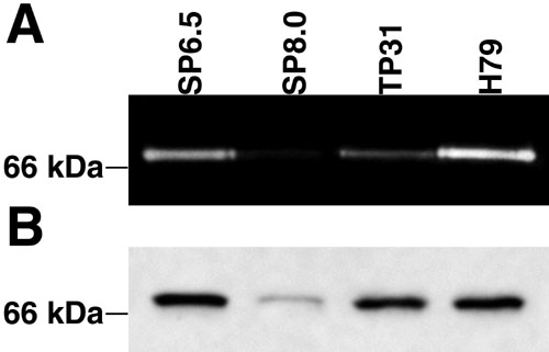
Figure 1. Zymographic and western blot analysis of MMP-2 from in vitro cultured uveal melanoma cell lines
A: Representative zymographic profile of the in vitro gelatinolytic activity in uveal melanoma cell lines. The in vitro gelatinolytic activity of MMP-2 was demonstrated in the serum-free conditioned media (25 μg each) from the SP6.5, SP8.0, TP31, and H79 uveal melanoma cell lines. The gelatinolytic activity observed at 72 kDa corresponds to proMMP-2. B: Western blot analysis of MMP-2 secreted by in vitro cultured uveal melanoma cell lines. For each sample, equal amounts of proteins (75 μg) were loaded on a 10% SDS-polyacrylamide gel prior to their transfer onto the membrane and further analysis with a monoclonal Ab directed against MMP-2. The position of the 66 kDa molecular mass marker is provided.
