![]() Figure 4 of
Gomez-Cabrero, Mol Vis 2005;
11:1071-1082.
Figure 4 of
Gomez-Cabrero, Mol Vis 2005;
11:1071-1082.
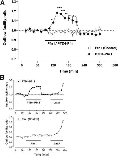
Figure 4. Effect of PTD4-Profilin I on trabecular outflow facility
A: Outflow facility ratio (normalized with respect to an initial 90 min baseline) is plotted against time. Solid dots correspond to perfusion with Dulbecco's Modified Eagle's Medium (DMEM) plus 2 μM PTD4-Pfn I (n=5) for 120 min (horizontal bar). Open dots correspond to perfusion with DMEM plus control Pfn I (n=6) for 120 min. A return to baseline conditions was seen after removing the fusion protein from the perfusion medium. The error bars represent the standard error of the mean. Statistically significant differences (one-way ANOVA with Bonferroni post hoc tests) were found between Pfn I and PTD4-Pfn I groups. The single asterisk indicates a p<0.05, the double asterisk indicates a p<0.01, and the triple asterisk indicates a p<0.001. B: Representative single experiments with PTD4-Pfn I (top) and control Pfn I (bottom) fusion proteins. In both experiments, 2 μM Latrunculin A (Lat A) was added at the end of the protocol as a positive control.
