![]() Figure 3 of
Houdart, Mol Vis 2005;
11:1061-1070.
Figure 3 of
Houdart, Mol Vis 2005;
11:1061-1070.
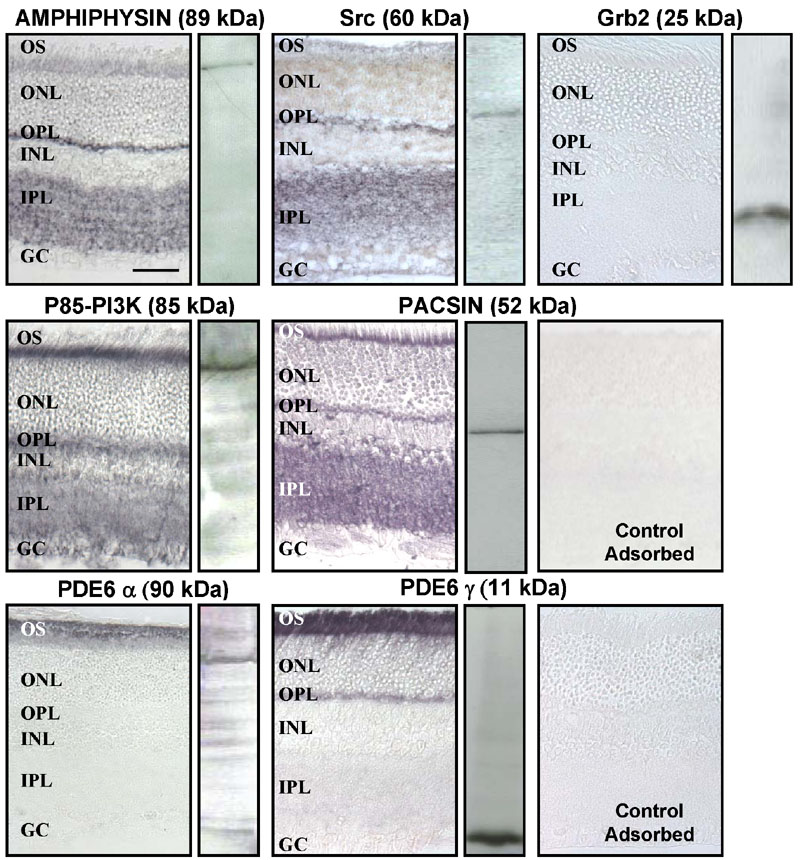
Figure 3. Immunodetection of SH3 domain-containing proteins and PDE6 subunits in the rat retina
Immunohistochemistry: eyes obtained in the middle of the day were fixed in Bouin's fixative and embedded in paraffin. Tissue sections (5 μm) were incubated overnight at 4 °C with the indicated antibodies (anti-AMPHIPHYSIN 1:500; anti-Src 1:100; anti-Grb2 1:100; anti-P85-PI3K 1:500; anti-PACSIN 1:250; control: anti-PACSIN 1:250 preadsorbed on rat cerebellum extracts; anti-PDE6α 1:20,000; anti-PDE6γ 1:10,000; control: anti-PDE6γ 1:10,000 preadsorbed on GST-PDE6γ fusion protein), then 1 h with a biotinylated secondary antibody (1:200). Immunoreactions were detected with the ABC complex and nickel-diaminobenzidin (the scale bar represents 50 μm). Western blots: retinas obtained in the middle of the day were homogenized in Laemmli sample buffer and analyzed by immunoblotting with the indicated antibodies, at the same concentrations as above, using the ECL method.