![]() Figure 2 of
Houdart, Mol Vis 2005;
11:1061-1070.
Figure 2 of
Houdart, Mol Vis 2005;
11:1061-1070.
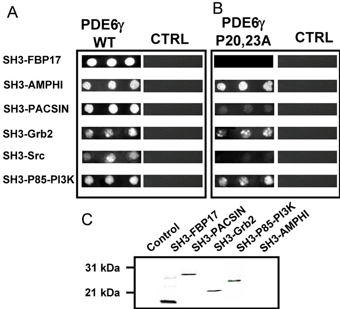
Figure 2. PDE6γ in vitro interaction with SH3 domains
A: Two-hybrid assay: L40 yeast expressing the wild type PDE6γ-rod bait (as lexADBD fusion) were transformed with the indicated SH3 domains cDNAs (as gal4AD fusions) and plated as triplicate drops on medium lacking histidine. Photographs were taken after 2 days growth at 28 °C. CTRL: L40 yeast expressing the irrelevant bait ERG19 was transformed and grown as above. B: L40 yeast expressing the PDE6γ-rod bait with the indicated proline mutations (as lexADBD fusion) or the irrelevant bait ERG19 (CTRL) were transformed and grown as above. C: GST pull-down assay: 35S-labeled SH3 domains were produced by in vitro transcription and translation and incubated overnight with glutathione-sepharose beads conjugated either to GST or to the GST-PDE6γ fusion proteins. After washes, proteins bound to the beads were eluted in Laemmli sample buffer and analyzed by SDS-PAGE and autoradiography.