![]() Figure 1 of
Keiser, Mol Vis 2005;
11:1052-1060.
Figure 1 of
Keiser, Mol Vis 2005;
11:1052-1060.
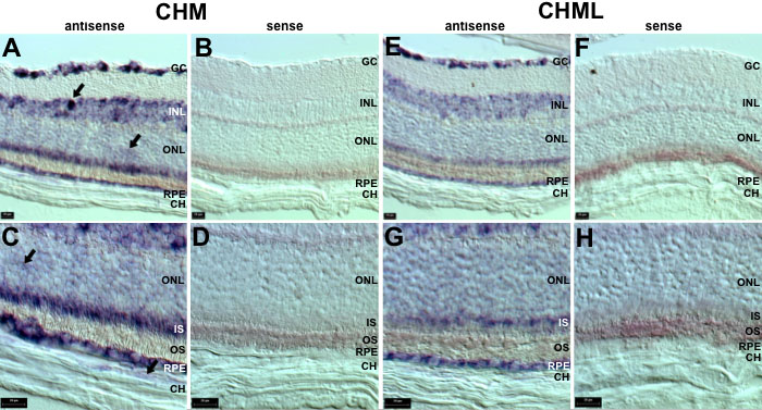
Figure 1. Localization of Chm and Chml transcripts in the retina
The results from in situ hybridization on 14 μm cryosections of adult (4 months) CD-1 mouse retina using sense and antisense RNA probes specific for Chm and Chml are shown. The sections were photographed under Nomarski differential interference contrast (DIC) microscopy using a 20x (A,B,E,F) or 40x (C,D,G,H) objective lens. A,C: The antisense probe was Chm. B,D: The sense probe was Chm. E,G: The antisense probe was Chml. F,H: The sense probe was Chml. Areas of strong Chm localization to the INL and localization to the ONL and choroid are indicated by the black arrows (A,C). Scale bars represent 20 μm. The ganglion cell layer (GC), inner nuclear layer (INL), outer nuclear layer (ONL), inner segments (IS), outer segments (OS), retinal pigment epithelium (RPE), and choroid (CH) are identified.