![]() Figure 2 of
Young, Mol Vis 2005;
11:1041-1051.
Figure 2 of
Young, Mol Vis 2005;
11:1041-1051.
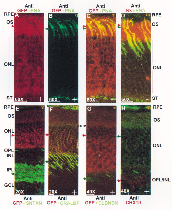
Figure 2. Activity of the rhodopsin kinase (Rk) enhancer/promoter in Rk-GFP transgenic eyes
The activity of the rhodopsin kinase (Rk) enhancer/promoter in Rk-GFP transgenic eyes is restricted to photoreceptors. Single channel (A,B) or merged (C-H) confocal micrographs of whole globe sections from Rk-GFP transgenic (A-C,E-H) or C57/BL6 (D) mice labeled with: polyclonal anti-GFP (red) and PNA (green) (A-C), polyclonal anti-Rk (red) and PNA (green) (D), polyclonal anti-GFP (red) and monoclonal anti-syntaxin (sntxn, green) (E), polyclonal anti-GFP (red) and monoclonal anti-CRALBP (green) (F), polyclonal anti-GFP (red) and monoclonal anti-calbindin (green) (G), and polyclonal anti-Chx10 (red) (H). Immunostaining in conjunction with a red fluorescent secondary antibody was used whenever possible to maximize GFP detection sensitivity (A-C,E-F). Direct GFP fluorescence was reliably detected as shown in panel H at higher gains and laser intensities over background in outer nuclear layer. Four 1 μm thick optical slices captured simultaneously in green (g) and red (r) channels were projected, viewed independently, or merged (m). Red and green arrows point to discrete areas of specific fluorescence and yellow arrows to sites of colocalized fluorescence (C,D). Magnification appears above a scale bar representing 20 μM. Images from animals with the GFP genotype are marked with a plus "+"; others are marked with a minus "-". The retinal pigment epithelium (RPE), outer segments (OS), outer nuclear layer (ONL), (OLM) outer limiting membrane, synaptic terminal (ST), outer plexiform layer (OPL), inner nuclear layer (INL), inner plexiform layer (IPL), and ganglion cell layer (GCL) are identified.