![]() Figure 3 of
Gupta, Mol Vis 2005;
11:1018-1040.
Figure 3 of
Gupta, Mol Vis 2005;
11:1018-1040.
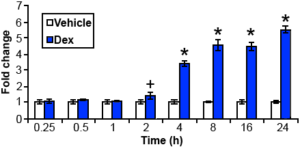
Figure 3. GRE activation in transfected HLE B-3 cells
Tenth passage HLE B-3 cells were co-transfected with pGRE.Luc and pRL-SV40 to normalize transfection efficiencies. Transfected cells were cultured in the presence or absence of dexamethasone for times indicated and luciferase activities were measured in each sample. Each time point was examined 2-5 times, each in triplicate, with similar results. Data are the mean of one experiment carried out in triplicate. The error bars represent the standard deviation (n=3). Values were significantly different (two tail, two sample t-test assuming unequal variances) from vehicle. The plus sign indicates p<0.02 and the asterisk indicates p<0.00002 when compared to the vehicle control at the same time.