![]() Figure 7 of
Andrieu-Soler, Mol Vis 2005;
11:1002-1011.
Figure 7 of
Andrieu-Soler, Mol Vis 2005;
11:1002-1011.
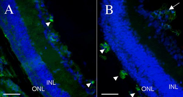
Figure 7. F4/80 immunohistochemistry on eye sections from treated and control PN28 rd1/rd1 mice
In order to evaluate cell infiltration, macrophages and infiltrating cells were labeled with F4/80. F4/80 immunostaining in green and DAPI staining in blue. Infiltrating macrophages (arrowheads) in rhGDNF-loaded microspheres treated eyes without microspheres aggregation (A) or with microspheres aggregates at the retina surface (B; white arrow). The outer nuclear layer (ONL) and inner nuclear layer (INL) are identified. Scale bars represent 100 μm.