![]() Figure 6 of
Andrieu-Soler, Mol Vis 2005;
11:1002-1011.
Figure 6 of
Andrieu-Soler, Mol Vis 2005;
11:1002-1011.
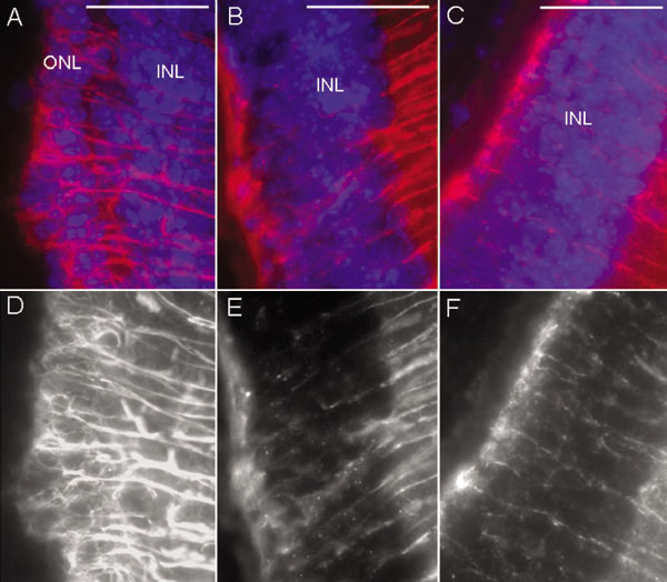
Figure 6. Glial reaction in the outer retina of rhGDNF treated eyes
GFAP immunostaining in red and DAPI staining in blue on sections from PN28 rd1/rd1 mouse: treated with rhGDNF-loaded microspheres (A) showing thick prolongation closely surrounding photoreceptors, treated with blank microspheres (B) or untreated (C) showing subretinal glial reaction and thin RMG prolongations. Corresponding black and white images of GFAP immunostaining are also shown in this figure (D-F). The outer nuclear layer (ONL) and inner nuclear layer (INL) are identified. Scale bars represent 100 μm.