![]() Figure 5 of
Andrieu-Soler, Mol Vis 2005;
11:1002-1011.
Figure 5 of
Andrieu-Soler, Mol Vis 2005;
11:1002-1011.
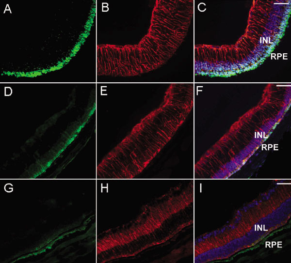
Figure 5. Rhodopsin and GFAP immunohistochemistry in eye sections from treated and control PN28 rd1/rd1 mice
Rods were identified by rho-4D2 immunostaining (in green) on sections from PN28 mice eyes treated with rhGDNF-loaded microspheres (A) or blank microspheres (D), or untreated (G). Glial cells were identified by GFAP immunostaining (in red) on sections from PN28 mice eyes treated with rhGDNF-loaded microspheres (B) or blank microspheres (E), or untreated (H). Rho-4D2, GFAP and nuclei (DAPI in blue) staining were combined on sections from PN28 mice eyes treated with rhGDNF-loaded microspheres (C) or blank microspheres (F), or untreated (I). The inner nuclear layer (INL) and retinal pigment epithelium (RPE) are identified. Scale bars represent 100 μm.