![]() Figure 4 of
Andrieu-Soler, Mol Vis 2005;
11:1002-1011.
Figure 4 of
Andrieu-Soler, Mol Vis 2005;
11:1002-1011.
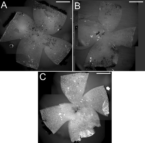
Figure 4. Rhodopsin immunohistochemistry on whole flat-mount retinas from treated and control PN28 rd1/rd1 mice
Representative pictures of flat-mounted retinas from untreated mouse (A), mouse treated with blank microspheres (B), and mouse treated with rhGDNF-loaded microspheres (C). D: Quantification of fluorescence resulting from rhodopsin immunohistochemitry (data are means of normalized fluorescence; error bars represent standard deviation) on whole flat-mount retinas from untreated, blank microspheres, and rhGDNF-loaded microspheres treated groups. A significant increase of immunofluorescence is observed in eyes treated with rhGDNF-loaded microspheres when compared to the eyes treated with blank microspheres (p=0.04) or to the untreated eyes (p=0.003). Scale bars represent 1 mm.