![]() Figure 3 of
Andrieu-Soler, Mol Vis 2005;
11:1002-1011.
Figure 3 of
Andrieu-Soler, Mol Vis 2005;
11:1002-1011.
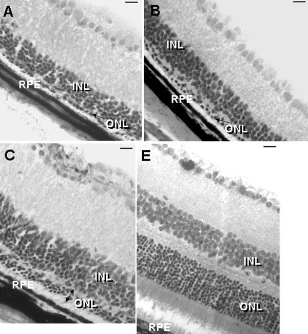
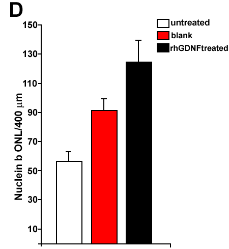
Figure 3. Eye sections and ONL cell counting at PN28
Hematoxylin-eosin stained retina sections of an rd1/rd1 untreated eye (A), eye treated with blank microspheres (B), eye treated with rhGDNF-loaded microspheres (C), and untreated wild type eye (E) at PN28. The outer nuclear layer (ONL), inner nuclear layer (INL), and retinal pigment epithelium (RPE) are identified. Arrows indicate the ONL. D: Counting of nuclei in the ONL shows a significant increase of nuclei in eyes treated with rhGDNF-loaded microspheres compared to untreated eyes (p=0.001) or eyes treated with blank microspheres (p=0.002). Scale bars represent 20 μm.