![]() Figure 2 of
Andrieu-Soler, Mol Vis 2005;
11:1002-1011.
Figure 2 of
Andrieu-Soler, Mol Vis 2005;
11:1002-1011.
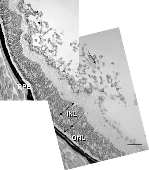
Figure 2. Aggregation of microspheres at the retinal surface as occurred in two of the injected eyes
Histology picture of retina section stained with hematoxylin-eosin shows that microspheres (arrows) are still present at 28 days and form aggregates at the retinal surface. Note that numerous photoreceptors have survived (double arrow showing ONL) at the site of microspheres accumulation. The outer nuclear layer (ONL), inner nuclear layer (INL), and retinal pigment epithelium (RPE) are identified. Scale bar represents 100 μm.