![]() Figure 2 of
Gu, Mol Vis 2005;
11:971-976.
Figure 2 of
Gu, Mol Vis 2005;
11:971-976.
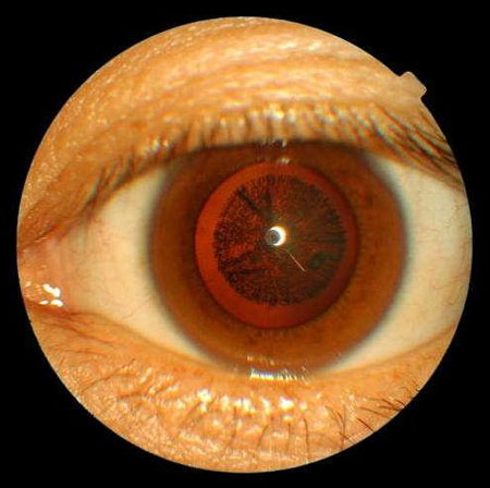
Figure 2. Frontal view photograph of the eye of the proband
Photograph of the anterior segment of the eye of the proband using a TOPCON Retinal camera TRC-NW6S. Opacities were located in the embryonal, fetal, and infantile nucleus of the lens while the cortex was transparent.