![]() Figure 5 of
Ghosh, Mol Vis 2005;
11:901-908.
Figure 5 of
Ghosh, Mol Vis 2005;
11:901-908.
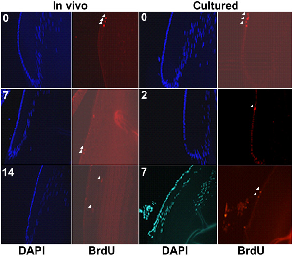
Figure 5. BrdU labeling of lens cells
BrdU labeling was used to compare the migration of lens epithelial cells and their differentiation into fibers in vivo and in lenses maintained in organ culture. For the in vivo study animals were euthanized at 0, 7, and 14 days after injection with BrdU. For the organ culture study, animals were euthanized 4 h after injection with BrdU, the lenses placed into modified TC-199 and removed from culture at 0, 2, and 7 days. All lenses were fixed in formalin and embedded in paraffin. Sections (6 μm) were stained using a monoclonal antibody specific for BrdU and with DAPI to visualize cell nuclei. The location of BrdU labeled nuclei are indicated by arrows demonstrating lack of migration in the cultured lenses. DAPI staining is shown to assist in orientation.