![]() Figure 5 of
Zhang, Mol Vis 2005;
11:887-895.
Figure 5 of
Zhang, Mol Vis 2005;
11:887-895.
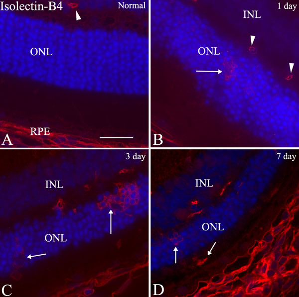
Figure 5. Isolectin-B4 labeling in light-exposed retinas
A: No isolectin-B4-positive cells were seen in the ONL in normal retina, except labeling of a small blood vessel (arrowhead) in the OPL. B: At 1 day, some isolectin-B4-positive round cells were seen in clusters in the ONL (arrow). C: At 3 day, more positive cells were seen in clusters in the ONL (arrows). D: At 7 day, the isolectin-B4-positive cells started to disappear from the ONL as the degenerative process subsided down. Scattered positive cell was seen in the subretinal space. The scale bar represents 20 μm.