![]() Figure 3 of
Zhang, Mol Vis 2005;
11:887-895.
Figure 3 of
Zhang, Mol Vis 2005;
11:887-895.
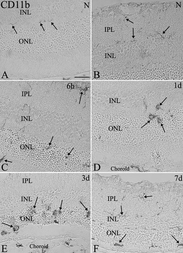
Figure 3. CD11b-positive cells in light-exposed retinas
A,B: In normal retinas, scattered CD11b-positive ramified microglial cells were noted in the outer plexiform layer (A, arrows), inner nuclear layer (INL), inner plexiform layer (IPL; B, arrows), and ganglion cell layer but not in the outer nuclear layer (ONL) or the subretinal space. C: Infiltration of CD11b-positive microglial cells in the ONL was observed at 6 h after photic injury. D,E: Marked infiltration of microglial cells in the ONL was seen at 1 day and 3 day after photic injury. F: By 7 day, a decrease of microglial cells was noted in the ONL but some remaining positive cells were seen in the subretinal space. The scale bar represents 20 μm.