![]() Figure 2 of
Zhang, Mol Vis 2005;
11:887-895.
Figure 2 of
Zhang, Mol Vis 2005;
11:887-895.
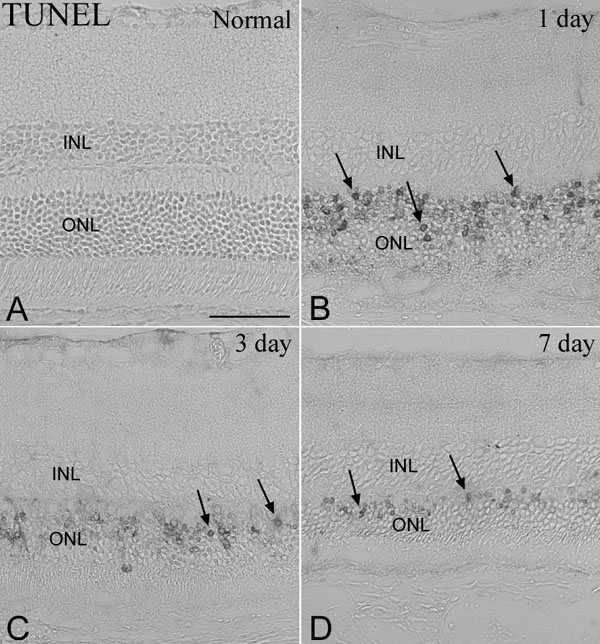
Figure 2. TUNEL in mouse retinas following photic injury
A: No TUNEL-positive cells were seen in the retinas of normal control Balb/cJ mice. B: TUNEL-positive cells (arrows) were most abundant 1 day after light exposure. C: TUNEL positive cells were less abundant 3 days after light exposure. D: Few TUNEL positive cells were seen 7 days after light exposure. The thickness of the ONL was markedly thinned at 3 and 7 days after light exposure. The scale bar represents 50 μm.