![]() Figure 1 of
Zhang, Mol Vis 2005;
11:887-895.
Figure 1 of
Zhang, Mol Vis 2005;
11:887-895.
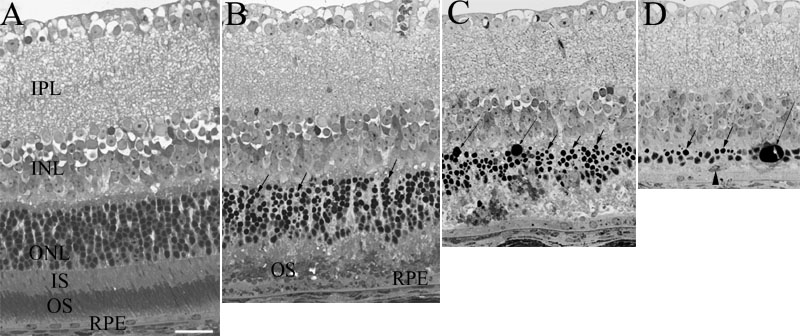
Figure 1. Photomicrographs from the equatorial regions of the superior quadrants of mouse retinas
A: Normal control retinas received the same exposure schedule except the experimental groups received an intense light exposure. Retinal pigment epithelium (RPE) was regularly arranged with oval nuclei. Photoreceptor inner and outer segments as well as photoreceptor nuclei were well aligned. B: One day after photic injury, the retinal pigment epithelium (RPE) cells were swollen with abundant phagocytosed photoreceptor outer segments. The inner and outer segments showed moderate disorganization. A large number of pyknotic nuclei appeared in the outer nuclear layer (ONL), especially in the inner part of the ONL (arrows). C: Three days after photic injury, the RPE cells remained swollen with round and prominent nuclei, the inner and outer segments were markedly thinned with severe disorganization. Photoreceptor cell loss was evident in the ONL with large number of pyknotic nuclei (short arrows). Large DNA globules, due to the coalescence of dying photoreceptor cell nuclei, were noted in the ONL (long arrows). D: Seven days following photic injury, the RPE cells seemed attenuated with subretinal infiltration of macrophage (arrowhead). The ONL thickness was remarkably thinned with little inner or outer segments. Many pyknotic photoreceptor nuclei were seen in the inner part of the ONL (short arrows). Large DNA globules were present (long arrow). The inner retinal layers appeared unremarkable throughout the observation period. The outer segments (OS), inner segments (IS), inner nuclear layer (INL), and inner plexiform layer (IPL) are identified. The scale bar represents 20 μm.